In our time, dying from radiation seems unlikely, and yet such a danger exists. Radiation protection requires certain precautions. The destruction of body cells that occurs when interacting with a radioactive object contributes to the development of many dangerous diseases.
Humans are exposed to background radiation quite often. The sun, marble, granite, radon gases - they all serve as sources, but their effect on the body is insignificant. Unfortunately, there are situations in which the risk of exposure to radiation is quite high and knowledge of the rules of radiation protection can save lives. Preventive measures consist of observing 3 postulates that will help minimize the harmful effects of radiation exposure : time, obstacles and distance.
Risk of radiation exposure
The process of spreading energy is called radiation. Infrared, ultraviolet, light, and ionizing radiation all fall under this category. From the standpoint of life protection and means of protection against radiation, the ionizing type is of keen interest. At high doses of radiation, the process of ionization of the substance promotes the formation of free radicals in cells that destroy the integrity of the cell membrane.
The radiation cannot be detected without the right equipment, making it a very dangerous enemy. Its penetration occurs through the respiratory and digestive systems and through the skin. It most actively affects cells that are in the process of dividing. This feature makes its impact especially harmful to a growing child’s body and requires careful protection from radiation.
In addition to the development of cancerous tumors, it causes the following diseases :
- infertility;
- mutations at the cellular level;
- leukemia;
- cataract;
- diarrhea;
- damage to various organs;
- blood diseases;
- radiation sickness.
It is necessary to distinguish between the concepts of radiation and radioactivity. The second is the property of substances to emit ionizing radiation, which requires the use of radiation protection equipment. The first is the ionizing radiation itself, wandering in open space and existing before being absorbed by another substance.
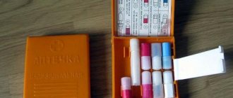
Scientific first aid kit
Thumbnail creation error: Cannot save thumbnail to destination
Yellow special medical aid kit
A medical aid kit designed for researchers conducting research activities in the Zone. The first aid kit contains drugs and products available in the “basic” version, which allows you to treat mechanical damage. This set also includes the drugs “Vikasol”, “Batilol”, S-naphthyzin and a set of drugs to prevent the development of radiation sickness.
Merchants do not sell this first aid kit.
Permissible radiation doses
The internal dose of radiation that enters the body along with water and food is the most dangerous. Unfortunately, decontamination methods used for external irradiation do not work here.
Radiation surrounds humans almost everywhere. For example, radon gas, which seeps in small volumes from the bowels of the earth and settles in the basements and first floors of buildings. Some household items, such as watches whose hands are treated with radium salt or a television, are also a source of radiation and require radiation protection. A classic example of exposure to radiation dose is the FLG procedure, which ideally should be performed annually. Products grown in a radioactive zone are also a dangerous source of contamination.
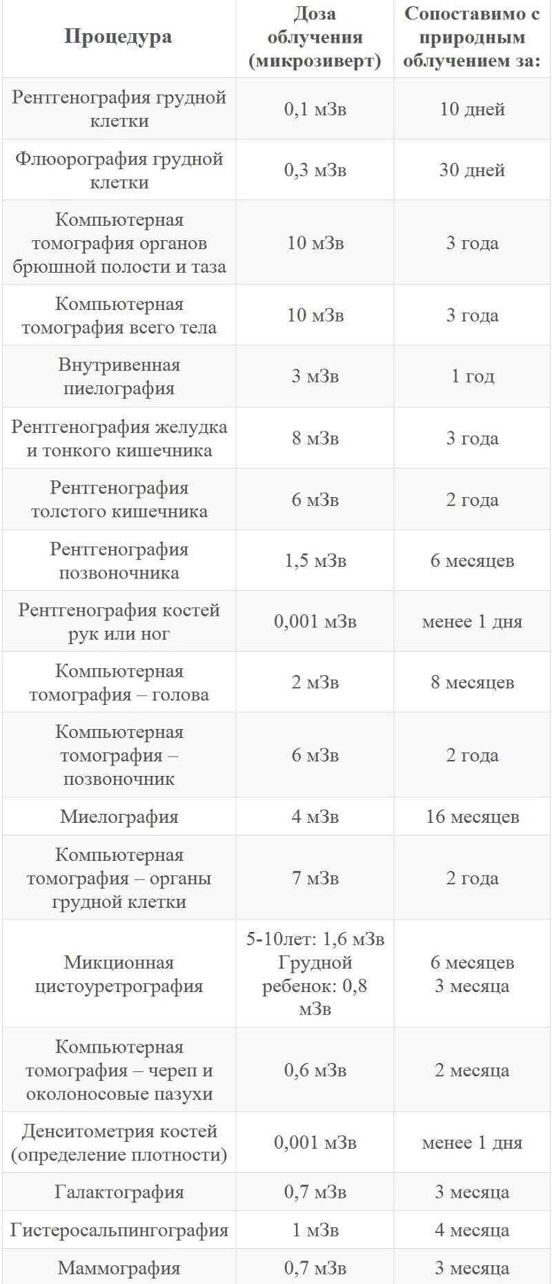
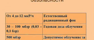
Any object is endowed with the ability to absorb radiation, and the human body is no exception. In this regard, the annual radiation dose for the majority of the population is set at 1 mSv. The radiation level is safe if it reaches no more than 0.5 mSv/h (microsievert per hour). The permissible exposure at an average rate is 0.2 mSv/h.
Anti-radiation drugs
Thumbnail creation error: Cannot save thumbnail to destination
Anti-radiation drugs
"Mexamine" is a drug that removes radionuclides from the body. It is not able to protect against the fact of receiving radiation, but is distributed throughout the Zone as the first means of combating radiation sickness. When used, peripheral blood vessels narrow and oxygen starvation is caused. This is what contributes to the prevention and treatment of radiation sickness. During the experiments, no systemic negative effects on the human body were observed, but isolated cases of nausea, headache, and even less frequently, vomiting were observed.
Methods of protection against radiation
When interacting with radioactive objects, all security methods are divided into 3 types:
- professional - for workers located in the affected area;
- medical – used in medical institutions;
- public – created with the aim of protecting the population.
In the social aspect, radiation protection means the use of barriers and compliance with time and distance rules in case of exceeding the permissible radiation dose.
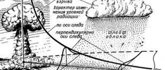
Based on the name, it is not difficult to guess that protection from radiation exposure consists of reducing the time spent near an object emitting radiation. Stay time should be minimized. It was this method that was used during the liquidation of the consequences of the accident at the Chernobyl nuclear power plant. To ensure protection from radiation, specialists were given only a few minutes to complete their task in the affected area. The radiation level rapidly decreases during the first hours after the explosion due to the decay of isotopes with a short life cycle. It then drops off quite slowly as the time comes for particles with long half-lives.
If you come into contact with an object that emits radioactive radiation and poses a health hazard, you should immediately move away from it. The impact power decreases as the distance between the person and the radiation source increases.
17.2. Anti-radiation drugsAnti-radiation drugs are drugs that increase the body’s resistance to the effects of ionizing radiation or reduce the severity of the clinical course of radiation sickness.
All personnel are provided with anti-radiation drugs. They are used by order of commanders (from the battalion commander and above) before crossing radioactive contamination zones or when operating in contaminated areas.
To prevent radiation sickness, troops are supplied with the drug RS-1, made in the form of tablets. The drug RS-1 is contained in a soldier’s individual first aid kit in two crimson pencil cases of 6 tablets each. This drug should be taken 30-40 minutes before entering an infected area or before exiting shelters into an infected area (6 tablets at a time). The protective effect of the drug lasts on average 6-7 hours. If the duration of irradiation is longer than the specified period, taking the drug is recommended
repeat in a similar manner from the second foam. After this, further use of the drug for 3 days is not advisable. Taking the drug RS-1 after irradiation does not have a protective effect. The degree of weakening of the effect of radioactive irradiation of RS-1 preparations is 1.3-1.5 times.
17.3. Antidotes
Antidotes are designed to neutralize toxic substances in the body. They play a major role in therapeutic and preventive care for those affected by poisoning! substances.
Antidotes are used by personnel independently when the first signs of poisonous substance damage appear or by order of the unit commander.
The antidote in the form of a solution is placed in a syringe tube for repeated or repeated use; administer: intramuscularly to the person affected by the toxic substance.
To administer the antidote from a syringe tube, you need to hold it in one hand, grab the ridges of the rim with the other and, rotating, push it towards the tube until it stops, so that the inner end of the needle pierces the m
tube membrane. Remove cap 4. Without touching the needle with your hands, insert it into the soft tissue of the front surface of the thigh or into the upper part of the buttock (you can through your uniform). Then, slowly squeezing body 2 with your fingers, insert its contents and, without unclenching your fingers, remove the needle.
To administer the antidote from an automatic, reusable syringe (
17.2), it is necessary to cock the spring 5 of the trigger mechanism 9, for which you take the trigger mechanism in your right hand, insert the fuse, rest the rod 7 against a solid object and compress the spring with a sharp push until it clicks. Remove plug 1 from cover 2 of the nozzle part and screw the cover onto the trigger mechanism. Take the syringe in your right hand and remove the safety lock with your left hand. Holding the syringe in your right hand, firmly attach the nozzle to the injection site of the antidote (thigh, shoulder or buttock) and, pressing the trigger handle, inject. After 6-8 seconds, remove the needle from the body. After removing the needle, put the fuse in place and disconnect the nozzle part. The trigger mechanism will be reusable.
Radiation protection equipment
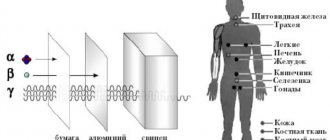
Radiation exposure is weakened by heavy substances, which act as a kind of protective shield. substances delay the effects of radiation :
- steel, 13 cm;
- water, 100 ml;
- brick, 40 cm;
- lead, 8 cm;
- loose soil, 90 cm;
- dense soil, 60 cm.
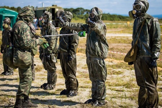
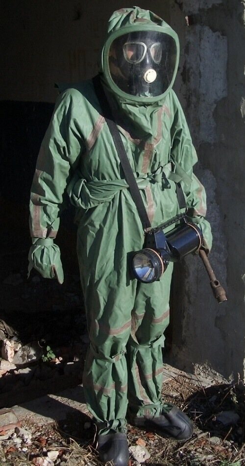
It is unsafe for people working in rooms with high background radiation to be present without the appropriate “equipment.” As methods of protection against radiation, there are specially designed screens that block ionizing radiation and a radiation suit.
For example, alpha radiation tends to affect only the skin when exposed externally. To ensure protection from radiation, you should use a respirator, gloves made of rubber, a polyethylene raincoat and cotton clothing.
Protecting yourself from beta radiation is a little more difficult. If the permissible radiation dose is exceeded, a glass or aluminum sheet shield and a gas mask will serve well. There is no need to study encyclopedias to understand how to build a shelter: just take shelter in the basement of a brick or concrete building.
The most difficult way to protect against radiation is when exposed to gamma radiation. The materials used to make the necessary uniforms - lead, tungsten, cast iron and steel - are quite expensive and have a high mass . How to make a shelter if it is not possible to determine the type of particles? Brick walls with interior decoration made of metal sheets and polyethylene will help protect you from the effects of any radiation dose.
RADIOPHARMACEUTICALS
Radiopharmaceuticals (RP)
— diagnostic or therapeutic agents containing radioactive nuclides. Diagnostic radiopharmaceuticals are used to study the anatomical and topographical state or assess the function of various organs and systems of the body. The effect of therapeutic radiopharmaceuticals on the pathological process is determined, in contrast to conventional biol drugs, by the action of radionuclide radiation (see Isotopes).
The use of radioactive tracers for scientific research was proposed in 1913 by D. Hevesy. In 1927, H. L. Blumgart and S. Weiss were the first to use a radioactive substance - radon (see) - for diagnostic purposes to determine the speed of blood flow. The systematic use of radionuclides in medicine began in the 40s. 20th century
The properties of a radiopharmaceutical are determined by the physical properties of the radionuclide and the chemical properties. compounds on the basis of which it was created. The main factors when choosing a radionuclide are the characteristics of its radiation (energy and yield of γ-quanta, half-life), as well as the possibilities and conditions of production. The half-life of a radionuclide (see Half-life) determines, along with other factors, the radiation dose created by the radiopharmaceutical for the patient and the possibility of using the drug for a specific study. According to Wagner (N. N. Wagner, 1966), the selected radionuclide must have a half-life equal to ln21t, where t is the research time after administration of the radiopharmaceutical. In this case, the relationship between the conditions for obtaining information during radioisotope research and the patient’s radiation dose is optimal. Since the absorbed dose is the sum of all types of radiation, radionuclides with a minimum yield of radiation not used for registration are preferred for diagnostic studies. Since the research methods are based on Ch. arr. on the registration of γ-quanta or characteristic X-ray radiation, this is best ensured by using radionuclides that decay through an isomeric transition (see Isomerism, atomic nuclei). The optimal limit for the energy of γ-radiation is 100-200 keV. At lower energy values, tissue absorption increases. With increasing energy, the efficiency of registration of radiation by the detector decreases and collimation becomes more complicated (see). An ideal radionuclide used to create a diagnostic radiopharmaceutical should have the following main characteristics: 1) one line of γ-quanta; 2) radiation energy 100-200 keV; 3) type of decay in the form of an isomeric transition or electron capture; 4) a half-life that does not differ significantly from the duration of the study.
The radionuclide used in therapeutic radiopharmaceuticals usually has not gamma, but beta radiation, which makes it possible to concentrate its action in the area of the pathological focus with minimal damage to surrounding tissues.
Chem. the compound (basic substance), on the basis of which a radiopharmaceutical is created, determines its biol, behavior in the body: the time of accumulation and retention in a particular organ, the rate of its elimination from the organ and the body. These indicators determine functional suitability, which is understood as the possibility of using a radiopharmaceutical for a given radiodiagnostic test or the possibility of therapeutic use. To give the radiopharmaceuticals the required distribution, various physiological and biochemical mechanisms are used, the main ones of which include participation in metabolic processes, phagocytosis, diffusion, physical-chemical. adsorption, cell sequestration, temporary capillary blockade, localization in a specific area of the body, antigen-antibody and enzyme-substrate reactions. In some cases, the mechanism that ensures this or that pattern of radiopharmaceutical distribution remains unclear. For therapeutic radiopharmaceuticals, the following chemicals are optimal. properties that can ensure selective accumulation of the drug only in the pathological focus.
A special group consists of diagnostic radiopharmaceuticals containing short-lived radionuclides obtained from generators. The generator is a system of genetically related two radionuclides - a long-lived parent and a short-lived daughter. The decay of the first of them leads to the formation of the second. The generator includes a column containing a particular sorbent with an adsorbed parent radionuclide, and a communications system for isolating the daughter radionuclide. The most widely used generator systems are 99Mo - 99mTc and 113Sn - 113mIn. The introduction in the mid-60s was of great importance. in wedge, practice 99mTc, the nuclear physical characteristics of which make it almost ideal for many radiodiagnostic methods. The use of preparations containing ultra-short-lived radionuclides (11C, 13N, 15O) produced in cyclotrons is developing. In this case, various inorganic compounds are used, for example. 11CO, 11CO2, 13NH3, 15O2, H215O, 13N2, 15CO2, as well as labeled amino acids, sugars, steroids, etc. These radionuclides decay with the release of positrons and the formation of two paired γ-quanta, scattering at an angle of 180°, which creates ideal conditions for emission tomography.
Quality control and quantitative characteristics of radiopharmaceuticals are established by the relevant national commissions (in the USSR - the Pharmacopoeial Committee M3 of the USSR). In the production of radiopharmaceuticals, various control methods are used: physical (determining radionuclide purity, volumetric and specific activity), chemical (determining radiochemical and chemical purity) and biological (determining sterility and pyrogenicity).
Radionuclide purity is the proportion of the total activity of the drug due to the required radionuclide. Radionuclide contaminants can increase the patient's radiation dose, reduce accuracy, and distort test results. Volume activity is the radionuclide content in 1 ml of the drug; is established taking into account the method of application and shelf life of the radiopharmaceutical. Specific activity - the content of a radionuclide per unit weight (mass) of the main substance; is determined by the possible influence of the amount of the latter on the biol, the behavior of the drug and its pharmacological (toxic) properties. These three parameters are monitored using radiometry (see) and spectrometry.
Radiochemical purity is the proportion of the radionuclide present in the radiopharmaceutical in the required chemical state. form. For example, if the radiochemical purity of 131I-hippuran is 98%, this means that 98% of the 131I in the preparation is associated with hippuran. Radiochemical impurities can significantly affect the reliability of the information received. In particular, 131I-hippuran with a radiochemical purity of at least 98% can be used to determine the effective blood flow of the kidneys. Chem. The purity of a drug is determined by the presence of foreign, unlabeled chemicals. In this case, special attention is paid to impurities of heavy metals. Control of radiochemical and chemical purity is carried out using chromatography (see), spectrophotometry (see), emission spectral analysis (see), etc.
Table 1. Diagnostic radiopharmaceuticals, the main research methods in which they are used, routes of administration and main properties
The use of diagnostic radiopharmaceuticals is based on the principle of radionuclide indication of chemicals. connections. Inclusion in the chemical composition. the radionuclide compound does not change its properties and makes it possible to monitor the distribution of radiopharmaceuticals in the body by external registration of radiation. Thus, radionuclides make it possible to directly study physiological and biochemical processes without disturbing their natural course, which is a fundamentally important advantage of radionuclide diagnostics. Its main methods are based on the principle of isotope dilution (determining the volume of circulating blood, plasma and red blood cells, water content in the body, etc.), on recording the distribution of radiopharmaceuticals in an organ or system (see Scanning, Scintigraphy), determining the amount of its accumulation in the organ or the lesion (diagnosis of the functional state of the thyroid gland with 131I, tumors - with 32P), registration of the kinetics of radiopharmaceuticals in an organ or tissue - renography (see Radioisotope renography), hepatography, determination of tissue blood flow and removal of radiopharmaceuticals from the body (diagnosis of impaired fat absorption in gastrointestinal tract). Table 1 provides a list of diagnostic radiopharmaceuticals, the main research methods in which they are used, methods of administration and basic properties.
Table 2. THERAPEUTIC RADIOPHARMACEUTICALS, MAIN INDICATIONS FOR USE, METHODS OF ADMINISTRATION AND QUANTITY OF ADMINISTRATION
The use of therapeutic radiopharmaceuticals is based on the destruction of tissues patol. focus by irradiating them. The main problem in this case is the concentration of the radiopharmaceutical in the area of the pathological focus, which is achieved either by its selective absorption, or, for example, by introducing it directly into the focus. Table 2 provides a list of therapeutic radiopharmaceuticals, the main indications for their use, methods of administration and the amount of the drug administered.
The use of radiopharmaceuticals is inevitably associated with radiation exposure to the patient's body, which theoretically represents the potential for somatic damage or genetic consequences. Calculation of risk values is difficult due to the sometimes very small amount of direct human data on which to base estimates, and therefore most calculations are based on extrapolation of risk at high doses. The tissue radiation doses created during the diagnostic use of radiopharmaceuticals in most cases amount to hundredths and tenths of a rad. In the Soviet Union, the USSR M3 approved “Radiation Safety Standards for Patients When Using Radioactive Substances for Diagnostic Purposes” are in effect, regulating the admissibility of a particular radiodiagnostic procedure depending on the purpose of the study. disease, age of the patient and generated radiation doses. The rule should be to reduce the radiation dose to such a minimum level that ensures satisfactory results of the study. The use of radiopharmaceuticals is contraindicated in pregnant and breastfeeding women, and in some cases in children under 16 years of age. In addition, some radiopharmaceuticals have specific contraindications.
The use of radiopharmaceuticals is permissible only in special radiodiagnostic departments in compliance with radiation safety measures for both medical personnel and the environment (see Radiation protection). The complex of radiation hygiene measures in the USSR is regulated by special legislative documents - “Radiation Safety Standards” (NSR) and “Basic Sanitary Rules for Working with Radioactive Substances and Other Sources of Ionizing Radiation.”
The main side effects when using radiopharmaceuticals are pyrogenic and allergic reactions, idiosyncrasy (see). Their frequency, according to various researchers, varies within very wide limits - from 0.4% to 15%.
See also Critical organ, Radioactive colloids, Radioactive drugs, Radioisotope diagnostics, Radioisotope research.
Bibliography:
Levin V.I. Obtaining radioactive isotopes, M., 1972; Parker R., Smith P. and Taylor D. Fundamentals of Nuclear Medicine, trans. from English, M., 1981; Atkins HL Radiopharmaceuticals, Physics reports, v. 21 cp 315, 1975; Blumgart H. La. Weiss S. Studies on the velocity of the blood flow, J. clin. Invest., v. 4, p. 15, 1927.
G. A. Zubovsky, N. F. Tarasov.